Search
(+)
cartoon
dealer
buy stock illustrations
Home
My rookie portfolio
📷 Upload yours 🤳
Find results that contain
all
of your keywords. Content filter is
on
. Search will return
best illustrations, stock vectors and clipart
.
Exclude
Make it so!
You have chosen to exclude "
" from your results.
Choose orientation:
Related commercial searches:
nasal
ocular
rhinosporidiosis
patient
3d
illustration
disease
caused
rhinosporidium
seeberi
parasite
Page
of 15
←
→
Order by:
relevancy of keywords
|
publishing date
|
most downloaded
Conjunctiva
Cartoons, Illustrations & Vector Stock Images
- 391 images
Nasal and ocular rhinosporidiosis in a patient, 3D illustration. A disease caused by Rhinosporidium seeberi parasite
Nasal rhinosporidiosis in a patient, 3D illustration. A disease caused by Rhinosporidium seeberi parasite
Ocular rhinosporidiosis in a patient, 3D illustration. A disease caused by Rhinosporidium seeberi parasite
The conjunctiva is a thin, clear membrane that protects your eye
Understanding Blepharitis and Conjunctivitis: A Detailed Close-Up of Eye Inflammation and Symptoms for
Conjunctivitis is an inflammation (swelling) or infection of the thin layer (conjunctiva)
The conjunctiva is a thin membrane that covers the inner surface of the eyelid and the white part of the eyeball
Eye layers
Optic part of retina.
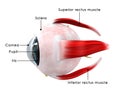
Eye oblique
Chalazion & sty
Conjunctivitis
Cone cells. Vector
Surface mucous cell
Anatomy of the human eye and visual apparatus
Photoreceptor cells in the retina of the eye
Pterygium
Human Eye
Anatomy of the Human Eye
Conjunctivitis
Next Page
←
↑
↓
→
See more images