Find results that contain all of your keywords. Content filter is on.
Search will return best illustrations, stock vectors and clipart.
Choose orientation:
Make it so!
You have chosen to exclude "" from your results.
Choose orientation:
Explore cartoons & images using related keywords:
skeleton
illustrationqua
illustrationquad
quaanatomy
muscle
quadriceps
femoris
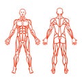
large composed distinct heads rectus superficial muscles middle originates male human anatomy triceps render brachii commonly upper arm humerus radial dorsri latissimus dorsi triangular located movements including shoulder spinous thoracic vertebrae thoracolumbar attaches intertubercular groove humanmuscle skeletion femour femur thighbone largest hip kneeMale Human Skeleton Femur Muscle Anatomy. 3d Render Royalty-Free Stock Photography
Designed by
Title
male human skeleton femur muscle anatomy. 3d render #275692073
Description


