Search
(+)
cartoon
dealer
buy stock illustrations
Home
My rookie portfolio
📷 Upload yours 🤳
Find results that contain
all
of your keywords. Content filter is
on
. Search will return
best illustrations, stock vectors and clipart
.
Exclude
Make it so!
You have chosen to exclude "
" from your results.
Choose orientation:
Explore cartoons & images using related keywords:
immunofluorescence
redistribution
myosin
actin
fibers
cell
direction
migration
microbiology
biochemistry
disease
microscopic
molecular
human
abstract
molecule
drug
bacterial
research
medical
laboratory
science
microscope
microorganism
pharmaceutical
scientific
experiment
medicine
bacterium
ai
generated
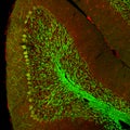
An Immunofluorescence Image Showing The Redistribution Of Myosin And Actin Fibers As A Cell Changes Direction During Its Stock Photo
Download
Designed by
Justlight
Title
An immunofluorescence image showing the redistribution of myosin and actin fibers as a cell changes direction during its #320564526
Description
An immunofluorescence image showing the redistribution of myosin and actin fibers as a cell changes direction during its migration. AI generated
Cerebellar folium, confocal image
An immunofluorescence image showcasing the unique distribution of lysosomes within a cell with some ed near the cell
An immunofluorescence microscopy image depicting the distribution of type I collagen in bone matrix, showing its fibrillar
Immunofluorescence image depicting the expression of various receptors on the surface of microglia marking their
An immunofluorescence image of apoptotic cells in a cell culture labeled with fluorescent markers to highlight the
An immunofluorescence image showing brightlycolored protein markers ed in the Golgi apparatus indicating active
An immunofluorescence image of a cell undergoing cellular respiration with bright spots indicating the active production
Immunofluorescence microscopic image of a cells cytoskeleton highlighting the close association between the NaK ATPase
An immunofluorescence image of glycoproteins labeled with a fluorescent dye on the surface of a red cell revealing their
An immunofluorescence image highlighting the key proteins involved in autophagosome formation such as LC3 and WIPI2
An immunofluorescence image highlighting the formation of phagolysosomes in various types of immune cells emphasizing
Immunofluorescence image highlighting the presence and distribution of different transcription factor proteins in a cell
An immunofluorescence image showing the localization of LC3 protein a key marker of autophagy in the cytoplasm
An immunofluorescence image highlighting the diverse modifications that can occur on histone proteins mediating their