Search
(+)
cartoon
dealer
buy stock illustrations
Home
My rookie portfolio
📷 Upload yours 🤳
Find results that contain
all
of your keywords. Content filter is
on
. Search will return
best illustrations, stock vectors and clipart
.
Exclude
Make it so!
You have chosen to exclude "
" from your results.
Choose orientation:
Explore cartoons & images using related keywords:
actin
antibody
antigen
biology
cell
cells
clinic
close
confocal
culture
cytosceleton
experiment
fibroblasts
fluorescence
human
immunology
laboratory
macro
mammalian
medical
medicine
microfilaments
microscope
microscopic
microscopy
nuclear
nucleus
research
science
scientific
sickness
skin
staining
tubulin
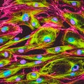
Confocal Microscopy Of Fibroblast Cells Stock Image
Download
Designed by
Vshivkova
Title
Confocal microscopy of fibroblast cells #75502129
Description
Real fluorescence microscopic view of human skin cells in culture. Actin filaments are labeled with green staining
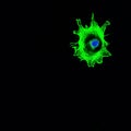
Confocal microscopy imaging of a single cancer cell invading
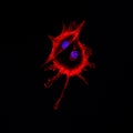
Confocal microscopy imaging of two cancer cells touching each ot
Confocal microscopy of fibroblast cells
A confocal microscopy image showcasing the constant assembly and disassembly of microtubule bundles during cell
A confocal microscopy image highlighting the role of intermediate filaments in supporting and stabilizing the cells
A confocal microscopy image revealing the location of ligandgated ion channels at the tips of dendritic spines
Confocal microscopy image capturing the localization of protein kinases in different cellular compartments
Confocal microscopy image of lipid rafts demonstrating their ability to move and fuse with each other
A confocal microscopy image of a cell undergoing stressinduced protein degradation with ubiquitin molecules forming
High magnification confocal microscopy image of endothelial cells expressing tight junction proteins aiding in the
Confocal microscopy image of a presynaptic terminal undergoing active recycling of synaptic vesicles after a burst of
A confocal microscopy image of a cell with bright red stains outlining the pathways through which endocytosis occurs
A confocal microscopy image of the dendrites of a neuron demonstrating the distribution of ligandgated ion channels
A confocal microscopy image showing the fusion of a secondary lysosome with a phagosome initiating the process of
A confocal microscopy image of epithelial tissue demonstrating the tight junctions as a band of fluorescence between the
A threedimensional reconstruction of a peripheral nerve created using multiple confocal microscopy images showing the